
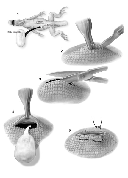
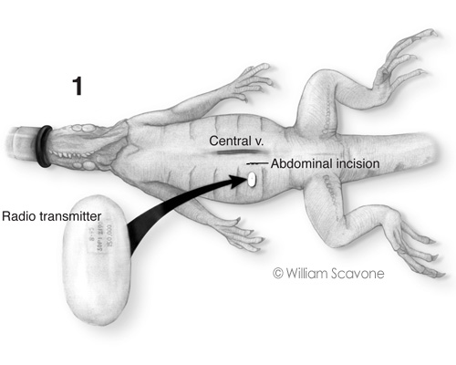
Surgical Implantation of Radio Transmitter into Iquana
This plate illustrates surgery to implant a radio transmitter into the abdominal cavities of an iguanas from the rainforest exhibit of the National Aquarium in Baltimore, Maryland.
The data transmitted by the device allowed researchers to map the iguana's movements through the exhibit while recording the animals' core temperature, thereby determining whether the habitat provided adequate warmth and exposure to sunlight, which was necessary to metabolize vitamin D and regulate calcium levels.
The surgery itself is fairly simple. Taking care to locate the incision well lateral of the central vein (1), the skin is tented with pick-ups to retract it from the viscera, and nicked with a #15 scalpel, passing through the abdominal wall and the adherent parietal peritoneum (2). While still tenting with the pickups, the tip of the scissors is inserted; maintaining ventral traction, the incision is extended, cutting through all layers, including the tough scales (3). Once the incision is slightly longer than the width of the transmitter, the implant is inserted into the abdomen. The central vein can be appreciated in this view (4). Last, the incision is closed with 2 horizontal mattress sutures, everting the edges of the wound (5). The transmitter can move freely about the abdomen.
Media: Airbrush dust (lamp black watercolor, film pencil, and ink on Laserline® board)
Scroll down to view a detail of the locator diagram.
© William Scavone. All Rights Reserved.