
|
|
||
|
|
|
|
|
||
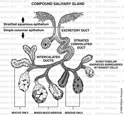
| Objective: To illustrate the various kinds of cells and the morphology of a compound salivary gland, clarifying the information for histology students. Used for the Johns Hopkins School of Medicine's Lecture Links website. Each part of the gland is shown sectioned at perpindicular angles so that the student will recognize the gland in histological sections.
References: Lecture Links Syllabus, Wheater's Functional Histology, Netter's Anatomy & Physiology Medium: Pen & ink, Adobe Photoshop Mode of Presentation/Audience: Web page / Medical students All images ©2001 William W. Scavone. All Rights Reserved. Images are not |